
Pilar Cossio
Max Planck Tandem Group Leader associated with the University of Antioquia (Colombia) and the Max Planck Institute of Biophysics (Germany)
Cryo-BIFE: Cryo-EM Bayesian Inference of Free Energy profiles
Date and time: March 3rd, 2021
Wednesday, at 8am PT / 11am ET / 4pm GMT / 5pm CET / Feb 18th midnight China
For the zoom and gather.town links, please join the One World Cryo-EM mailing list.
Note that we no longer use the zoom registration system, so old zoom links may not work. The new links will be sent via the mailing list.
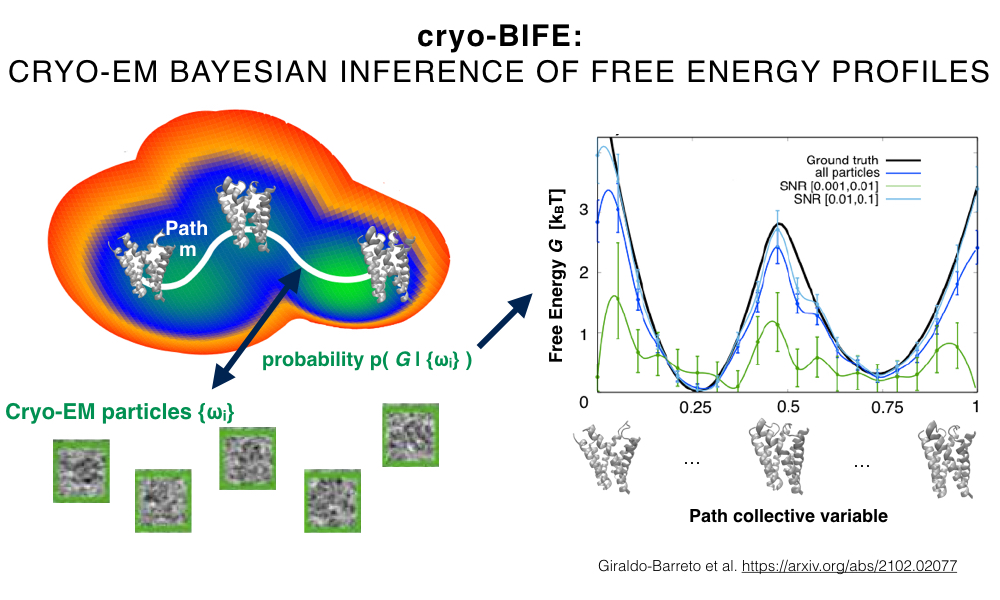
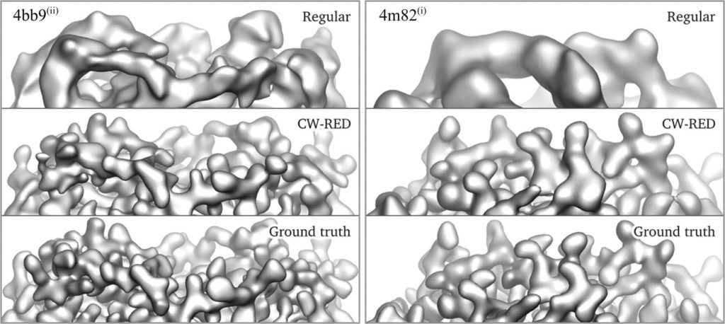
Cryo-electron microscopy (cryo-EM) extracts single-particle density projections of individual biolmolecules. Although cryo-EM is widely used for 3D reconstruction, due to its single-particle nature, it has the potential to provide information about a biomolecule’s conformational variability and underlying free energy landscape. However, treating cryo-EM as a single molecule technique is challenging because of the low signal-to-noise ratio in the individual particles. In this work, we developed the cryo-BIFE method, a Bayesian framework that uses a path collective variable to extract free energy profiles and their uncertainties from cryo-EM images. We tested the framework over several systems, finding that for realistic cryo-EM environments, and relevant biomolecular systems, it is possible to recover the underlying free energy.