
Edward H Egelman
Professor, Biochemistry and Molecular Genetics, University of Virginia
Cryo-EM and Helical Polymers: A Natural Affinity
Date and time: 9/23/2020 Wednesday, at 8am PT / 11am ET/ 4pm BST / 5pm CEST / 11pm China
Link: please join our mailing list at https://mailman.yale.edu/mailman/listinfo/ow_cryoem
It was over fifty years ago that the first 3D reconstruction from electron microscopy was published (DeRosier and Klug, 1968), and this happened to be a helical phage tail. It was no accident that this was a helical specimen, and not just because helical polymers are so abundant in biology. From one point of view helical objects are the simplest to reconstruct: a single image of a helical filament may provide all of the information needed for a 3D reconstruction. Fourier-Bessel methods of reconstruction dominated the field for the next 30 years, but now real-space approaches have become the standard. With the advent of direct-electron detectors, reaching a near-atomic resolution for helical polymers has become the standard, rather than the exception. However, problems in determining the correct helical symmetry can persist even when one reaches a resolution of ~ 5 Å. I will discuss how the best approach to determining helical symmetry involves an averaged power spectrum from the images, and explain why the power spectrum of an averaged image (such as using Class2D) is not the same as the averaged power spectrum. In addition, the highly anisotropic environment present in thin films prior to vitrification, the compressional forces associated with these thin films, and fluid flow with associated shear, can all impact the structure of the filaments being examined. I will discuss these problems and potential solutions while giving an overview of some of our own efforts involving everything from viruses infecting hosts living in nearly boiling acid to microbial nanowires, with stops along the way at bacterial and archaeal flagellar filaments and bacterial and archaeal pili.


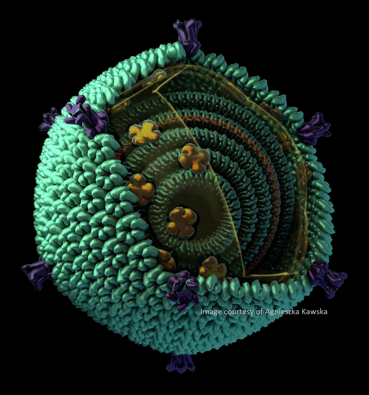
Images courtesy of Agnieszka Kawska.