2020/12/02: Alberto Bartesaghi: CryoET – high-speed or high-resolution? BISECT tackles both

Alberto Bartesaghi

Alberto Bartesaghi

Associate Professor of Computer Science, Biochemistry and Electrical and Computer Engineering, Duke University

CryoET – high-speed or high-resolution? BISECT tackles both

Date and time: Dec 2nd, 2020 Wednesday, at 8am PT / 11am ET / 4pm GMT / 5pm CET / Dec 3rd midnight China

For the zoom and gather.town links, please join the One World Cryo-EM mailing list.
Note that we no longer use the zoom registration system, so old zoom links may not work. The new links will be sent via the mailing list.

Tomographic reconstruction of cryo-preserved specimens followed by extraction and averaging of sub-volumes has been successfully used to determine the structure of macromolecules in their native environment.
Eliminating biochemical isolation steps required by other techniques, this method opens up the cell to in-situ structural studies. Delays introduced during mechanical navigation of the specimen and stage tilting, however, significantly slow down data collection thus limiting its practical value. Here, I present BISECT (beam image-shift electron cryo-tomography), a new protocol to accelerate tilt-series acquisition without sacrificing resolution. I also describe improvements to our constrained single particle tomography (CSPT) framework, leading to higher resolution reconstructions determined by sub-volume averaging.
For validation, we collected tilt-series from a low molecular weight target (~300kDa) using BISECT and processed them using CSPT to obtain a 3.6 Å resolution map where density for side chains is clearly resolved. These advances bring cryo-ET a step closer to becoming a high-throughput tool for in-situ structure determination at near-atomic resolution.

2020/11/18: Jane Richardson: The Challenge of Model Validation & Correction for 2.5-4Å CryoEM

Jane Shelby Richardson

James B. Duke Professor of Medicine
Professor of Biochemistry
Duke University

The Challenge of Model Validation & Correction for 2.5-4Å CryoEM

Date and time: Nov 18th, 2020 Wednesday, at 8am PT / 11am ET / 4pm GMT / 5pm CET / Nov 19th midnight China

Registration: https://yale.zoom.us/meeting/register/tJApcu6grD4iGdFaQsDVdpJEaGD1oE-3VOjB
If you registered to the previous talk your zoom link will still work.

Please register to our mailing list to receive updates. Please use institution/company email.

New! gather.town breakout room before the talk. Please register to our mailing list to receive the link.

2020/11/4: Bridget Carragher: CryoEM: Challenges and Opportunities

Bridget Carragher
Director, Simons Electron Microscopy Center,
New York Structural Biology Center, NY, NY, USA

CryoEM: Challenges and Opportunities

Date and time: Nov 4th, 2020 Wednesday, at 8am PT / 11am ET / 4pm BST / 5pm CET / Nov 5th midnight China

Bridget will talk about challenges and opportunities in cryoEM; then, we will invite participants to add their perspective. What are the things that existing algorithms and software cannot do currently? What would make experimental work better? What do we expect or hope to achieve in the coming years?

Advance registration is required: https://yale.zoom.us/meeting/register/tJApcu6grD4iGdFaQsDVdpJEaGD1oE-3VOjB

2020/10/21: Alex Noble: Neural network particle picking and denoising in cryoEM with Topaz

Alex Noble
National Resource for Automated Molecular Microscopy, Simons Electron Microscopy Center,
New York Structural Biology Center, NY, NY, USA

Neural network particle picking and denoising in cryoEM with Topaz

Date and time: 10/21/2020 Wednesday, at 8am PT / 11am ET/ 4pm BST / 5pm CEST / 11pm China

Single particle cryoEM projects are often hampered by low SNR particle views, which are missed by most particle pickers or severely de-prioritized causing junk to be preferentially picked. Moreover, for non-globular, small, asymmetric, and aggregated proteins, picking and centering such particles becomes critical. To solve these issues and more, we present Topaz particle picking using a novel positive-unlabelled framework and Topaz-Denoise using the Noise2Noise framework. We show that Topaz and Topaz-Denoise significantly increase the number of real particles picked, enable conventionally difficult projects, significantly decrease classification bias, and increase collection efficiency. We show the first in-depth analysis of pre-trained 2D and 3D denoising models for cryoEM and cryoET, which remove the characteristic sheets of noise in cryoEM micrographs of proteins and cryoET cellular tomograms. We also will highlight Topaz pre-trained picking models. In the past several months, Topaz has been used by numerous labs around the world to enable and optimize single particle cryoEM projects, including several SARS-CoV2 projects. These recent projects and more will be highlighted to show the unique and timely power of Topaz. We will also highlight Topaz integration into the most popular cryoEM suites: Relion, CryoSPARC, Scipion, and Appion.

2020/10/7: Jasenko Zivanov: Bayesian Particle-Polishing in Detail

Jasenko Zivanov
MRC Laboratory of Molecular Biology, Cambridge

Bayesian Particle-Polishing in Detail

Date and time: 10/7/2020 Wednesday, at 8am PT / 11am ET/ 4pm BST / 5pm CEST / 11pm China


Registration required: https://yale.zoom.us/meeting/register/tJApcu6grD4iGdFaQsDVdpJEaGD1oE-3VOjB
Please join our mailing list at https://mailman.yale.edu/mailman/listinfo/ow_cryoem

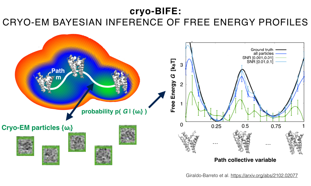
The beam-induced motion-correction method informally known as Bayesian Polishing will be discussed in detail. The method has been made available to the public in 2019 as part of Relion-3 and has been widely adopted since. The talk will focus on the Bayesian formulation of the problem and on the use of Gaussian processes to model our prior assumptions on the movement of particles embedded in the ice. A more recent adaptation of the method to estimate 3D motion in tomographic samples will also be briefly presented.

Zivanov, Jasenko, Takanori Nakane, and Sjors HW Scheres. “A Bayesian approach to beam-induced motion correction in cryo-EM single-particle analysis.” IUCrJ 6.1 (2019): 5-17.

2020/09/23: Edward H Egelman: Cryo-EM and Helical Polymers: A Natural Affinity

Egelman

Edward H Egelman
Professor, Biochemistry and Molecular Genetics, University of Virginia

Cryo-EM and Helical Polymers: A Natural Affinity

Date and time: 9/23/2020 Wednesday, at 8am PT / 11am ET/ 4pm BST / 5pm CEST / 11pm China

Link: please join our mailing list at https://mailman.yale.edu/mailman/listinfo/ow_cryoem

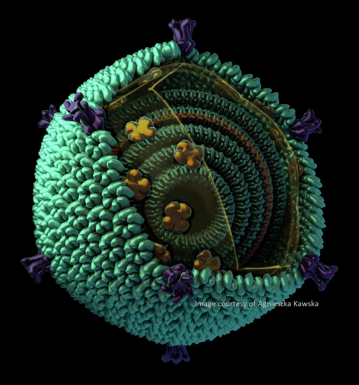
It was over fifty years ago that the first 3D reconstruction from electron microscopy was published (DeRosier and Klug, 1968), and this happened to be a helical phage tail. It was no accident that this was a helical specimen, and not just because helical polymers are so abundant in biology. From one point of view helical objects are the simplest to reconstruct: a single image of a helical filament may provide all of the information needed for a 3D reconstruction. Fourier-Bessel methods of reconstruction dominated the field for the next 30 years, but now real-space approaches have become the standard. With the advent of direct-electron detectors, reaching a near-atomic resolution for helical polymers has become the standard, rather than the exception. However, problems in determining the correct helical symmetry can persist even when one reaches a resolution of ~ 5 Å. I will discuss how the best approach to determining helical symmetry involves an averaged power spectrum from the images, and explain why the power spectrum of an averaged image (such as using Class2D) is not the same as the averaged power spectrum. In addition, the highly anisotropic environment present in thin films prior to vitrification, the compressional forces associated with these thin films, and fluid flow with associated shear, can all impact the structure of the filaments being examined. I will discuss these problems and potential solutions while giving an overview of some of our own efforts involving everything from viruses infecting hosts living in nearly boiling acid to microbial nanowires, with stops along the way at bacterial and archaeal flagellar filaments and bacterial and archaeal pili.

Images courtesy of Agnieszka Kawska.